MICROBIOLOGY

Microbiology is the study of living organisms of microscopic size, including bacteria, fungi (mould and yeast), algae, protozoa and viruses.
Some milestones in microbiological history
- Antonie van Leeuwenhoek, 1632 – 1723, a self-taught Dutchman, constructed the microscope with which he could observe bacteria. Leeuwenhoek has been called the “father of microscopy”.
- Louis Pasteur, 1822 – 1895, a French chemist, invented the heat treatment method that is now called pasteurization.
- Robert Koch, 1843 – 1910, a German physician and 1905 Nobel Prize winner for medicine, discovered pathogenic (disease-producing) bacteria such as the tubercle bacillus and cholera bacterium. In addition, he devised ingeniously simple methods to enable safe study of these organisms.
- Alexander Fleming, 1881 – 1955, a British microbiologist, professor and 1945 Nobel Prize winner for medicine, discovered penicillin (1929), which is effective against many bacteria, but not tuberculosis.
- Selman A. Waksman, 1888 – 1973, an American mycologist, microbiologist and 1952 Nobel Prize winner for medicine, discovered streptomycin, which is effective against many bacteria including tuberculosis.
Microbiology is the study of organisms that are so small, they can only be seen with the aid of a microscope. Size is measured in micrometer (µm). 1 µm = 0.001 mm
Microorganisms in nature
Microorganisms are found everywhere – in the atmosphere, in water, on plants, animals and in soil. Because they break down organic material, they play an important role in the cycle of nature.
Microorganisms occur most abundantly where they find food, moisture, and a temperature suitable for their growth. Since the conditions that favour the survival and growth of many microorganisms are those under which people normally live, it is inevitable that we live among a multitude of microbes.
Listed below are the key characteristics of the different groups of microorganisms.
Protozoa
- Unicellular, small aquatic organisms
- Can ingest solid food particles
- Ultimately become food for fish and larger animals
- Generally not food spoilage organisms
- A few protozoa are food-borne pathogens, transmitted by water
- Some protozoa are pathogens, transmitted by insects
Algae
- Uni- or multicellular organisms, frequently found in water
- Contain chlorophyll and are photosynthetic
- Used as food supplement and in pharmaceutical products
- Some are source of agar for microbiological media
- Some produce toxic substances
- Generally not food spoilage organisms
Yeasts
- Unicellular, oval or round in shape
- Frequently found in many environments: soil, plants and fruits
- Used as food supplement and in the production of alcoholic beverages
- Food spoilage organisms, especially of high-acid foods
Moulds
- Multicellular with many distinctive features
- Always found in soil, but can also be found in water and air
- Responsible for the decomposition of many materials
- Useful for industrial production of many chemicals, including penicillin
- Can cause diseases in humans, animals, and plants
- Food spoilage organisms, especially of high-acid foods
Bacteria
- A highly variable group of microorganisms
- Found in almost all environments
- Some cause diseases, others perform important roles in the natural recycling of elements, and thus contribute to soil fertility
- Useful in industries for the manufacture of valuable compounds
- Some spoil foods and others are used in the manufacture of foods
Viruses
- Can only reproduce in living cells i.e. are parasites
Parasites = microorganisms living on living animals and plants
Biotechnology
The concept of “biotechnology” is a fairly recently coined word for techniques utilising biological processes. Biotechnology has a history that predates the modern scientific disciplines of microbiology, biochemistry and process technology by thousands of years.
Until the end of the nineteenth century, these processes were associated with food, and above all with the preservation of food.
Microbial processes still play a prominent part in the food industry, but biotechnology in the modern sense is largely associated with industrial utilisation of the properties of living cells or components of cells to obtain production of various products, such as enzymes, hormones and certain vaccines. To accomplish this, it is necessary to utilise knowledge of the biosciences – biochemistry, microbiology, cell biology, molecular biology and immunology – as well as the technologies of apparatus design, process engineering, separation techniques, and analytical methods.
This chapter deals mainly with microorganisms relevant to milk and milk processing, but specific viruses called bacteriophages are also described. Bacteriophages cause serious problems in the manufacture of products where microorganisms are needed for the development of flavour, texture and other characteristics.
Bacteria
Bacteria are unicellular organisms that normally multiply by binary fission, i.e. splitting in two. Bacteria are classified partly by their appearance. However, to be able to see bacteria, they must be studied under a microscope at a magnification of about 1,000 times.
Bacteria may also be stained and the most widely used method of staining bacteria was introduced by the Danish bacteriologist Gram and is called Gram staining. Bacteria are divided into two main groups according to their Gram stain characteristics: Gram negatives are red and Gram positives are blue. This is due to differences in the bacterial envelope and gives the two groups of bacteria different properties.
Morphology of bacteria
Morphology is the study of the form of bacteria. This covers morphological features such as shape, size, cell structure, motility (ability to move in a liquid), and spore and capsule formation.
Shape of bacteria
Bacteria come in many different shapes. However, three main characteristic shapes can be distinguished: spherical-, rod- and spiral-shaped.
The spherical bacteria are named cocci; when arranged in pairs they are called diplococci (Figure 4.2); and when arranged in chains they are called streptococci (b). Cocci can also be arranged in irregular clusters like grapes (c) and in groups of four or as cubic arrangement (d).
Rod-shaped bacteria vary in both length and thickness. Some of them also form chains in the same way as many Bacillus species (Figure 4.3). The word bacilli means small rods.
Spiral bacteria can be of variable length and thickness, and the number of turns also differs. Spiral bacteria with many turns are among the largest bacteria, up to 20 μm are common.
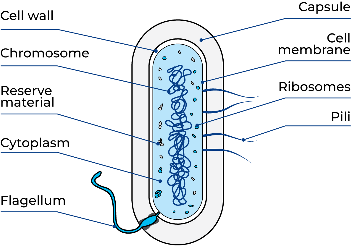
Cell wall - gives the cell its specific shape
Cell membrane - active transport of nutrients and metabolites
Cytoplasm - place of production of cell constituents
Chromosome - carrier of genetic information
Cell structure and function of bacteria
The smallest basic unit of life, both functionally and structurally, is a cell, and bacteria have the simplest cell organisation. The bacterial cell is prokaryotic. All other cells, including fungi, algae, protozoa, plant and animal cells, are eukaryotic. The minimum number of structural components in a bacterial cell is relatively small. The cell wall is the only rigid structural component. It gives the cell its shape. Bacteria with a thick cell wall are Gram-positive and bacteria with a thin cell wall are Gram-negative. The cell membrane determines what should be inside and outside of the cell. Only water-soluble substances can be transferred through the cell membrane. Thus, no exchange between the cell and its environment nor growth can take place if water is not present. The Gram-negative bacteria also have a second membrane, made of lipopolysaccharides, outside the cell wall. This can be measured if the bacteria have already been killed, which indicates enzymatic activity (itself sometimes too low to measure).
The contents of the interior of the bacterial cell are dispersed into two recognisable parts: the cytoplasm and the chromosome. The chromosome carries the genetic information in its DNA (deoxyribonucleic acid). In principle, a bacterium contains only one chromosome, i.e. one molecule of DNA. Its information is transformed into enzymes in the cytoplasm. In electron microscope pictures, the cytoplasm most often appears granular. The actual sources of production of cell components are the ribosomes, which consist of proteins and RNA (ribonucleic acid). In addition to the recognisable structural components (Figure 4.4), there are many solutes in the cytoplasm: break down products, vitamins, co-factors and building blocks for the synthesis of cell components.
Motility of bacteria
Some cocci and many bacilli are capable of moving in a liquid nutrient medium. They propel themselves with the help of flagella, which are long, stiff, thread-like appendages growing out of the cytoplasmic membrane (Figure 4.5). The length and number of flagella vary from one type of bacterium to another.
The bacteria generally move at speeds of between one and ten times their own length per second. The cholera bacterium is probably one of the fastest; it can travel 30 times its length per second.
Prokaryotic are all bacteria
- Simple cell organisation
- No membrane around the genetic material, which is one DNA molecule
Eukaryotic are fungi, algae, protozoa, plant and animal cells
- Cell organisation complex
- Membrane around the genetic material (several DNA molecules) i.e. a true nucleus exists
Spore formation
Only a few genera of bacteria form spores: Bacillus and Clostridium are the most well-known. The spore is a form of protection against adverse conditions, e.g. heat, disinfectants, dry conditions or lack of nutrients. Some of the different types of spore formations are illustrated in Figure 4.6.
When the parent cell forms a spore, it may retain its original shape, or it may swell in the middle or at one end, depending on where the endospore is located. During spore formation, the vegetative part of the bacteria cell dies. The cell eventually dissolves, and the spore is released.
The spore germinates back into a vegetative cell and starts reproduction when conditions become favourable again.
Spores are resting cells with no metabolism and thus cannot multiply. They can survive for years in dry air, and they are more resistant than bacteria to chemical sterilants, antibiotics, drying and ultraviolet light. They are also resistant to heat: 20 – 30 minutes in hot water or steam at 120 °C will generally destroy.
Simple cell organisation
No membrane around the genetic material, which is one DNA molecule
Eukaryotic are fungi, algae, protozoa, plant and animal cells
Cell organisation complex
Membrane around the genetic material (several DNA molecule) i.e. a true nucleus exists
Capsule formation
Some bacilli and cocci are surrounded by a capsule of strongly developed mucus. This makes them rather resistant to dry conditions. The growth of such bacteria in milk makes it viscous and slimy. In both cases, this phenomenon gives ropy milk.
Growth factors for bacteria
Nutrients
Bacteria require certain nutrients for their growth. The need for nutrients varies widely among different bacteria. The main sources of food are organic compounds, e.g. proteins, fats and carbohydrates. In addition, small amounts of trace elements and vitamins are necessary for growth.
As well as material for cell formation, organic matter also contains the necessary energy. Such matter must be soluble in water and have a low molecular weight, i.e. it must be broken down into very small molecules in order to be able to pass through the cytoplasmic membrane and be metabolised by the bacterium. Consequently, bacteria need access to water.
Some microorganisms lack the ability to release enzymes for breaking down substances outside the cell. They have to utilise breakdown products created by other microorganisms. Such a relationship is called symbiosis when both parties benefit from it. When one organism produces substances that have an inhibiting effect on other organisms, the process is called antibiosis.
Permanent union between organisms, each of which depends for its existence on the other
Antibiosis
An antagonistic situation where one organism produces substances which inhibit the growth of other organisms
Water activity
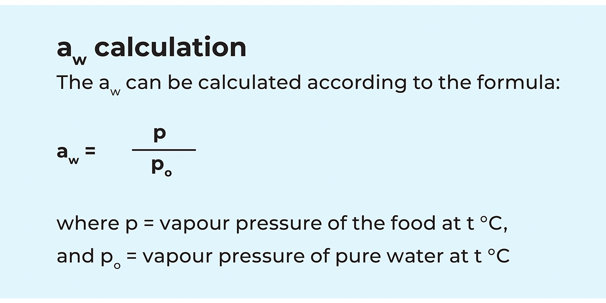
The growth and metabolism of microorganisms demand the presence of water in an available form. The most useful measurement of the availability of water is water activity, aw. The aw in a food may be reduced by increasing the concentration of solutes in the aqueous phase of the food, either by removing water or by adding solutes. Some water molecules are oriented around the solute molecules, and others become absorbed into insoluble food constituents. In both instances, the water becomes less available to enter into chemical reactions.
Dehydration is a method of food preservation that depends on the reduction of aw by water removal. In salting and sugaring, the addition of a solute lowers the aw and preserves the food. A small reduction in aw often has sufficient effect to preserve a food when combined with other factors.
Definitions of water activity
Dehydration is a method of food preservation that depends on the reduction of aw by water removal. In salting and sugaring, the addition of a solute lowers the aw and preserves the food. A small reduction in aw often has sufficient effect to preserve a food when combined with other factors.
Effect of water activity on growth
Dehydration is a method of food preservation that depends on the reduction of aw by water removal. In salting and sugaring, the addition of a solute lowers the aw and preserves the food. A small reduction in aw often has sufficient effect to preserve a food when combined with other factors.
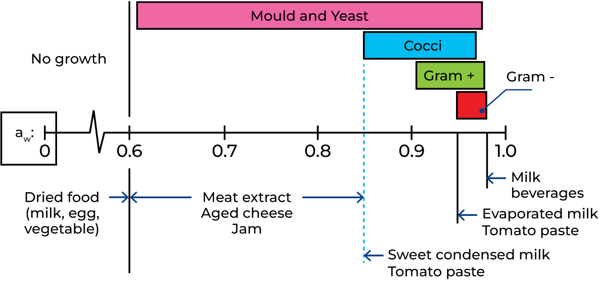
Most microorganisms relevant to food have their optimum growth rate at an aw of 0.98 or higher. This is also the range for aw of milk and beverages in general. Several gram-negative bacteria are the most competitive due to growth rate, i.e. if present, they will dominate the microbial flora. In milk, for example, a gram-negative infection often hides an infection of gram-positive bacteria.
At an aw between 0.98 and 0.93 the gram-positive bacteria dominate, i.e. Lactobacillus, Bacillus and Micrococcus, but a few tolerant coliforms may be present. Food-borne bacterial pathogens (Salmonella, C. botulinum and C. perfringens) are prevented from multiplication. In contrast, Staphylococcus grow at 50% of their maximum growth rate at aw = 0.94. Within this range, spoilage of low-acid food by fungi is possible and they can compete with bacterial spoilage.
At an aw between 0.93 and 0.85 possible spoilage organisms are cocci, moulds and yeasts. The only bacterial pathogen growing within this range is S. aureus. Intermediate moisture food is designed to have an aw < 0.85 in order to inhibit S. aureus.
At an aw between 0.85 and 0.60 a few moulds and yeasts can cause spoilage. Common foods in this range are jams and jellies preserved with a high concentration of sugar. There is no production of mycotoxin possible in this aw range. No microorganisms can grow at an aw < 0.60. Such food is safe from further microbial spoilage. It should be emphasised, however, that due to previous history, such food might contain viable microorganisms including pathogens and/or toxins.
Temperature
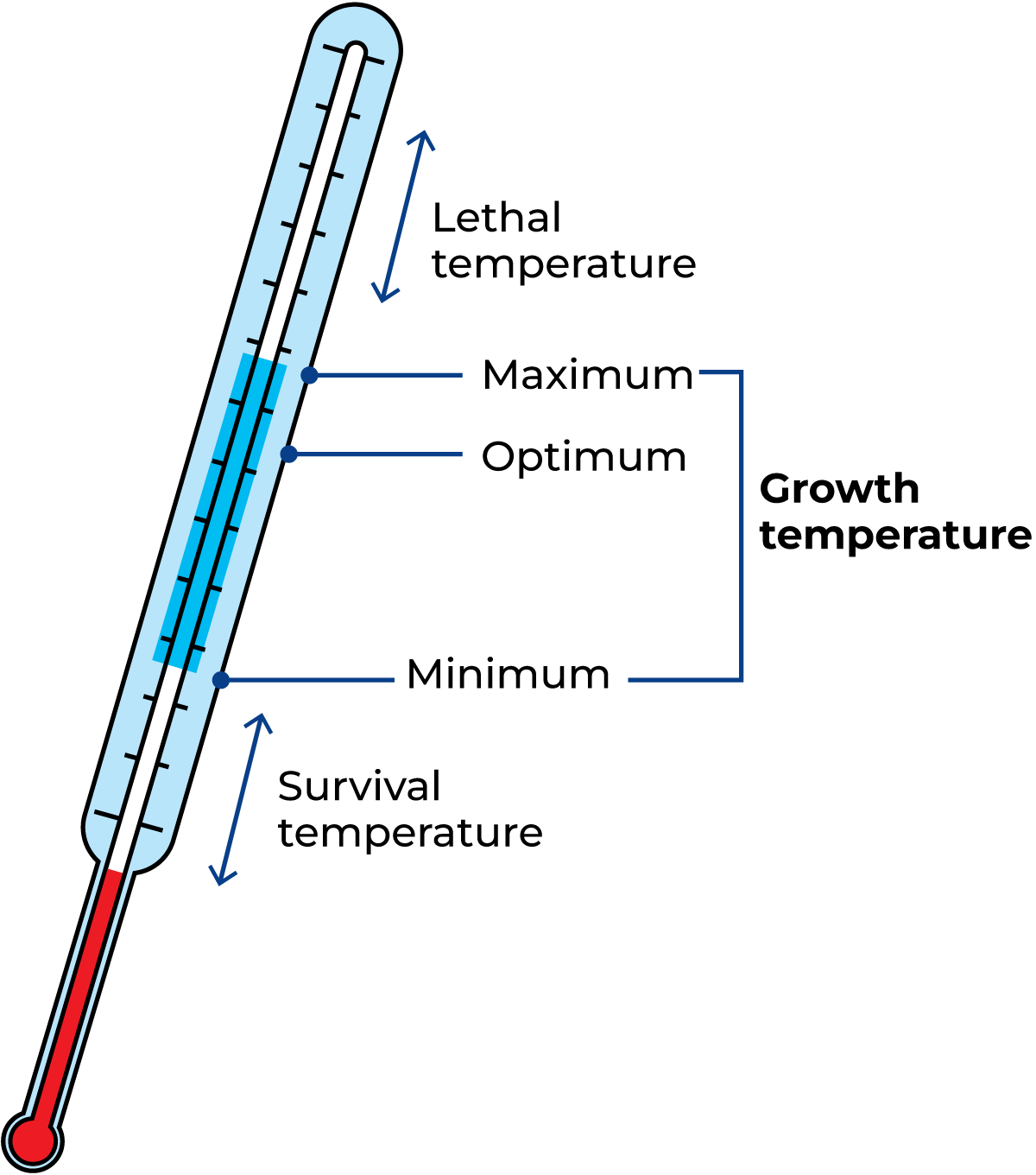
Temperature is the greatest single factor affecting growth, multiplication and food deterioration (Figure 4.8). Bacteria can only develop within certain temperature limits, which vary from one species to another. In principle, bacteria can grow at temperatures between the freezing point of water and the temperature at which the protein in the cytoplasm coagulates. Somewhere between the maximum and minimum temperatures, i.e. the upper and lower limits lies the optimum temperature. This is the temperature at which the bacterial strain multiplies most vigorously.
Temperatures below the minimum cause growth to stop, but do not kill the bacteria. The life functions of bacteria cease almost completely at a temperature close to the freezing point of water. As the cells have a high content of water, they will freeze at this temperature. When this happens, the bacteria can no longer absorb nutrients through the cell membranes.
If the temperature is increased above the maximum, the bacteria are quickly killed by heat. Most cells die within a few seconds of being exposed to 70 °C, but some bacteria survive heating to 80 °C for five minutes, even though they do not form spores.
It takes much more heat to kill bacterial spores, and dry heat is less effective than moist heat. Treatment with steam at 120 °C for 30 minutes ensures the destruction of all spores, but in dry heat, the bacteria must be kept at 160 °C for two hours to guarantee destruction of spores.
Classification by temperature
Bacteria can be divided into the following categories (Figure 4.9) according to their preferred temperature range: Psychrophilic (cold-loving) bacteria grow well at 0 °C with an optimum temperature of about 12 – 15 °C and a maximum of below 20 °C.
Psychrotrophic (cold-tolerant) bacteria are mesophilic strains that can multiply at commercial refrigeration temperatures with an optimum temperature of about 20 – 30 °C.
Mesophilic bacteria have a minimum temperature of about 10 °C, generally an optimum of 30 – 35 °C and a maximum of about 50 °C. Without a doubt, this is the most common range for bacterial growth. Approximately 90% of all bacteria can grow in this temperature interval.
Thermophilic (heat-loving) bacteria have optimum growth temperatures of 55 – 65 °C, with a minimum temperature of about 37 °C and a maximum of around 70 °C. The psychrotrophic bacteria are of particular interest to the dairy industry because microbiological activity in farm milk and market milk usually takes place at a temperature of 7 °C or below.
Oxygen
Many microorganisms need free oxygen to oxidise their food in order to produce energy and support their life processes. Complete oxidation of organic compounds forms CO2 and water. Many microorganisms can utilise air at atmospheric pressure, and these are called aerobic microorganisms. Other types obtain energy from their food without the need for free oxygen, and these are called anaerobic microorganisms.
There are some bacteria that consume free oxygen if it is present, but which can grow in the absence of free oxygen. Such bacteria are called facultatively anaerobic. Anaerobic and facultatively anaerobic bacteria generally obtain their energy by fermentation of organic compounds. Chemically, this is an incomplete oxidation, whereby organic waste products are formed, e.g. lactic acid from lactose, Table 4.2. As most organisms obtain their oxygen from the air, i.e. they are aerobic, removal of oxygen/air is a means of controlling or preventing their growth.
Examples of this are vacuum packing, gas packing and the use of materials acting as an air barrier. Anaerobic bacteria die if exposed to atmospheric oxygen for any length of time.
Light
Light is only essential for photosynthetic cells, which capture energy from the light. Microorganisms, including most bacteria, tend to be killed when exposed to direct sunlight. The ultraviolet part of the sunlight causes chemical changes in the DNA and cell protein.
Ultraviolet light is often used to disinfect atmospheres in starter rooms. However, it is not used to disinfect food, as chemical changes may also take place in the food.
pH – effect of acidity on growth
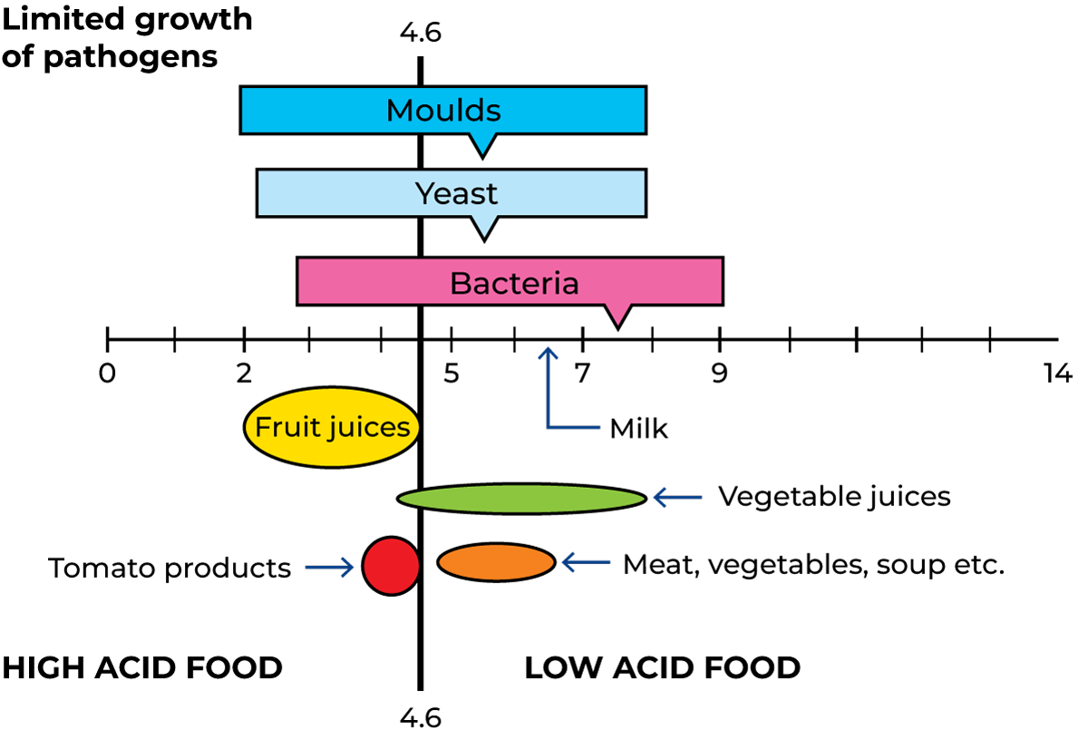
Most natural environments have pH values between 5 and 9, and the most common microorganisms are optimised for growth in this range. Most moulds and yeasts grow best in slightly acidic media, around pH 5 to 6, while optimum conditions for bacteria are neutral or slightly alkaline environments.
Fresh milk normally has a pH between 6.6 and 6.8. Moulds and yeasts generally grow well at pH as low as 3, or even pH 2. Most non-processed foods have a pH slightly below neutral, i.e. they are low-acid foods. Fruit juices are generally high-acid foods (Figure 4.10).
Multiplication of bacteria
Bacteria normally multiply by binary fission. In Figure 4.11, multiplication is shown graphically. Each individual cell grows and after reaching a critical size, it divides into two identical cells. The type of cell arrangement, which results in a characteristic cell grouping, is usually constant for a given species of bacteria. Cell grouping can take the forms of chains, pairs and clumps. This characteristic is therefore used in the description of different species.
Rate of multiplication
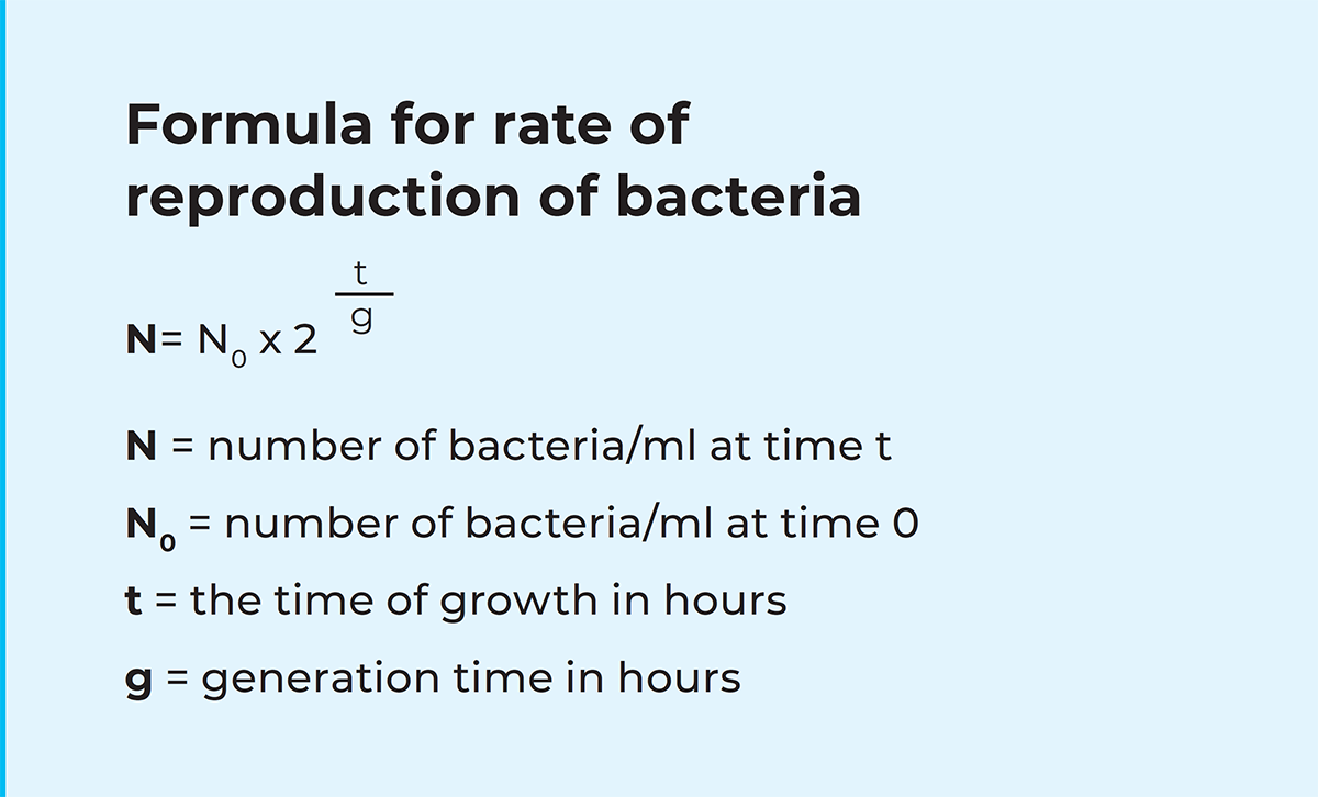
In favourable conditions, the multiplication of bacteria can occur at intervals of 20 – 30 minutes. The rate of multiplication can be calculated from the formula shown below. With a generation time of 0.5 hours, one bacterium/ml of milk will become about one million bacteria/ml within 10 hours. Under optimal conditions in food, 100 million – 1 billion bacteria/ml can be formed. At that stage, the growth rate will be inhibited by a lack of nutrients and the accumulation of toxic metabolic waste products. Reproduction finally stops, and large numbers of bacteria die. In reality, unfavourable conditions, such as low storage temperature or low pH will limit or delay the growth of bacteria in food.
Growth curve of bacteria
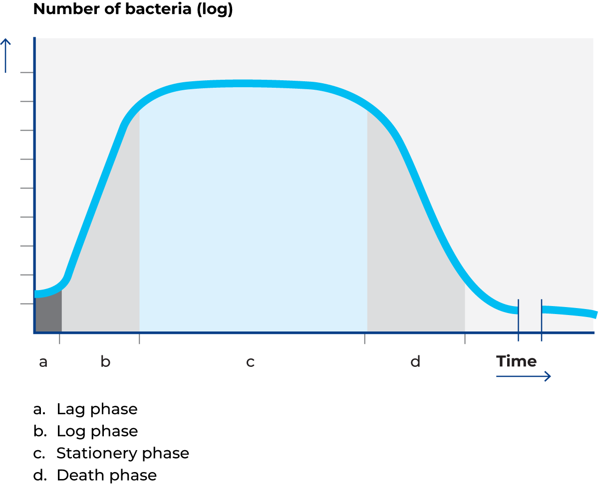
Figure 4.12 shows a curve of the growth of bacteria transferred to a substrate by inoculation. There is usually some delay before the bacteria start to reproduce, as they must first adapt to the new environment. This phase of development (a) is called the lag phase. The lag phase may also be because the culture needs to recover due to, for example, having been stored at a low temperature prior to inoculation.
The length of the lag phase varies according to how much the bacteria were inhibited at the moment of inoculation. After the lag phase, the bacteria begin to multiply logarithmically. This phase (b) is called the log phase or the exponential phase.
After some time, toxic metabolic waste products accumulate in the culture. The rate of multiplication will therefore subsequently slow down, while at the same time, bacteria are constantly dying so that a state of equilibrium is reached between the death of some cells and the formation of new ones. This phase (c) is called the stationary phase.
In the next phase (d), the formation of new cells ceases completely, and the existing cells gradually die off. Finally, the culture is almost extinct. This is called the death phase.
The shape of the curve, i.e. the length of the various phases and the gradient of the curve in each phase, varies with temperature, food supply and other
Biochemical activity
Due to biochemical activity, microorganisms can spoil food and cause diseases in animals and plants. Some microorganisms have biochemical activities that are used in food processes, i.e. the manufacture of cheese, yoghurt, butter, etc. The activity of a specific microorganism is governed by the enzymes it possesses, as these determine what it can feed on and break down, and consequently, what end-products it produces.
There are many biochemical and enzymatic systems in microorganisms. The following systems are the major ones concerned with milk and milk products. They can be subdivided into which constituent they break down and their effects.
The most important biochemical and enzymatic systems of bacteria in milk products are those responsible for the following effects:
- Breakdown of carbohydrates
- Breakdown of protein
- Breakdown of fat
- Breakdown of lecithin
- Production of colour
- Production of mucus or slime
- Production of odours
- Reduction of oxygen
- Diseases
Breakdown of carbohydrates
Carbohydrates or sugars have the formula (CH2O)n and were originally identified as hydrates of carbon. There are mono-, di- and polysaccharides. Polysaccharides are constituted of long chains of one or several sugars, for example, cellulose, starch and chitin. The enzymes of the microorganism determine which carbohydrates they can break down, and to what extent. In milk, hydrolysis of the disaccharide lactose causes the breakdown into glucose and galactose.
They can be completely degraded to carbon dioxide and water (oxidative metabolism), but in most cases, fermentation occurs.
Fermentation usually results in various products such as organic acids (lactic acid, butyric acid, etc.), alcohols (ethyl alcohol, butyl alcohol, etc.) and gases (carbon dioxide, hydrogen, etc.), Table 4.3.
The most important forms of fermentation due to activity of microorganisms are:
- Alcoholic fermentation of carbohydrates to alcohol and gas. One example is the breakdown of lactose into ethyl alcohol and carbon dioxide. Alcoholic fermentation usually takes place under anaerobic conditions and is mainly induced by yeasts.
- Lactic acid homofermentation of lactose, with lactic acid as the only end product. This reaction is used in the manufacture of cheese, yoghurt and other acidified products.
- Lactic acid heterofermentation of lactose, which produces lactic acid, acetic acid, carbon dioxide and ethyl alcohol.
- Coliform fermentation (mixed acid and butanediol) of lactose, which creates a wide variety of end products such as lactic acid, acetic acid, succinic acid, formic acid, butanediol, ethyl alcohol, carbon dioxide and hydrogen.
- Butyric acid fermentation under strict anaerobic conditions by some species of Clostridium bacteria. In butyric fermentation, lactose is broken down to butyric acid, carbon dioxide, hydrogen and, in some cases, butyl alcohol.
As a general rule, carbohydrate fermentations in milk result in the production of acid (souring) and, in some cases, gas (depending on the organisms).
Breakdown of carbohydrates by:
- Hydrolysis
- Alcoholic fermentation
- Lactic acid fermentation
- Coliform type fermentation
- Butyric acid fermentation
Breakdown of protein
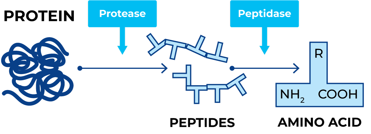
The process in which protein is broken down is called proteolysis. Proteases such as rennin, pepsin and trypsin are the main enzymes involved in the process. These enzymes degrade proteins into peptides, which are then degraded by various peptidases into smaller peptides and free amino acids. Amino acids can be reutilised for protein synthesis by the cell; however, they can also be broken down oxidatively or fermentatively.
Proteins and their constituent amino acids have a wide combination of chemical elements and contain carbon, hydrogen, oxygen, sulphur, nitrogen and phosphorus. Breakdown of protein therefore results in a much larger range of acids, alcohols, gases (hydrogen, carbon dioxide, hydrogen sulphide and ammonia) and other compounds. Breakdown of protein nearly always results in ammonia, which is alkaline and has a strong odour, Figure 4.13.
Three amino acids, cystine, cysteine and methionine, contain sulphur and result in hydrogen sulphide, which also gives off a strong smell like rotten eggs.
Breakdown of protein in liquid milk takes place in two major stages called peptonisation and consists of:
- Curdling (sweet as opposed to sour) or clotting of the milk by renin-like enzymes. This fault in milk is called sweet curdling, a defect which is common in pasteurized milk that is stored warm.
- Proteolysis of the protein, resulting in the production of ammonia, which is alkaline.
The degree of free amino acids and ammonia in cheese gives an indication of its age and maturity as proteolysis progresses. Blue, or mould-ripened cheese has rapid proteolysis, resulting in the production of large amounts of ammonia.
Breakdown of fat
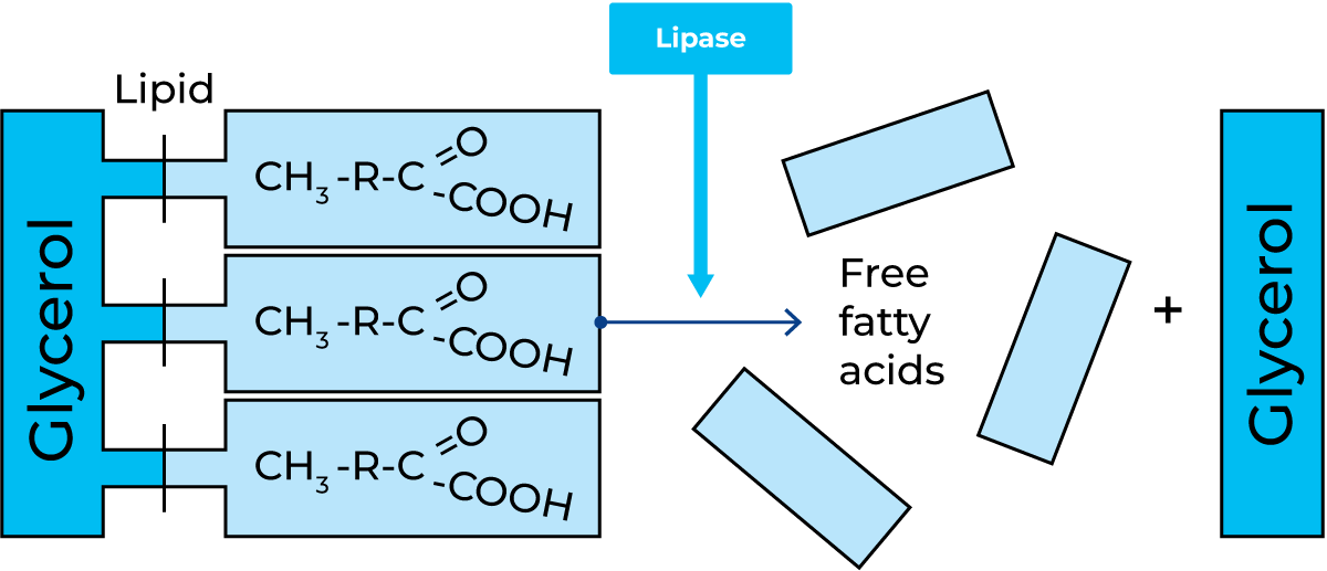
The process in which fat is broken down by enzymes is called lipolysis. Lipase is the main enzyme involved in this process. During lipolysis, the fat is hydrolysed to glycerol and one, two or three separate fatty acids, Figure 4.14. Some of the fatty acids are volatile and give off strong smells. One example is butyric acid, which gives the characteristic rancid taste.
Pure fat cannot be broken down by microorganisms, but fat in water emulsions, or fat in contact with water, is broken down by many microorganisms. Water is essential for enzymatic split. Milk fat in the form of butter and cream is a water emulsion and contains protein, carbohydrates, minerals, etc., which sometimes makes it even more susceptible to enzymatic breakdown.
Many bacteria and moulds that break down proteins also break down fat oxidatively.
Breakdown of lecithin
Lecithin, the phospholipid contained in the membranes around the fat globules, is a chemical combination of glycerol, two fatty acids, phosphoric acid and choline, an organic alkali. Strains of Bacillus cereus produce enzymes, lecithinases, which hydrolyse the lecithin into diglyceride and phosphorylcholine. The membranes of the fat globules are split, resulting in an unstable fat emulsion often seen in the form of flocs or lumps floating on the surface of the milk or cream. This fault in milk or cream is called bitty or broken cream.
Further breakdown of the choline into trimethyl amine will result in a fishy smell and taste.
Pigment and colour production
The process of colour production is called chromogenesis and the organism causing the production is referred to as chromogenic.
This process of metabolism is a feature of certain microorganisms. It is greater in some foods than others and takes place only at ambient or lower temperatures. Aerobic conditions are also favourable for chromogenesis.
There are two types of pigment:
- Endo-pigment, which stays in the cell
- Exo-pigment, which diffuses out of the cell into the surrounding food
There are three basic colour groups:
- Carotenoids, (yellow, green, cream or golden)
- Anthocyanins, (red)
- Melanins, (brown or black)
The name of an organism often refers to the colour it produces. Example: Staphylococcus aureus = the golden Staphylococcus.
for example:
Albus = white
Luteus = yellow
Citreus = citrus yellow
Roseus = pink or red
Aureus = golden
Violaceum = violet
Nigra = black or brown
Mucus production
A number of bacteria produce a mucus (or slime) of polysaccharides, which dramatically increase viscosity, as they are highly water-soluble and dissolve in the medium. This is utilised in certain cultured products such as yoghurt and “långfil”, a Swedish ropy milk.
Odour production
Some organisms produce strong odours or smells. Below is a list of some organisms and their associated smells:
- Moulds – musty
- Actinomycetes – earthy
- Yeasts – yeasty
- Pseudomonadaceae – fruity/fishy
- Coliforms – manure
- Lactococcus lactis var. maltigenes – malty
Pathogens in raw milk
Some microorganisms may cause food poisoning (pathogenic microorganisms, Table 4.4), either by intoxication and/or infection. Intoxication implies the production of poisons in the food prior to its consumption. Infection means the establishment, active growth, and multiplication of such microorganisms in the human body. Often rather large numbers are needed to cause an infection, but sometimes, as in the case of Escherichia coli O157:H7 (EHEC), the MID (minimum infection dose) may be as small as one bacterium.
Study of bacteria
Bacteria occur in nature in extremely large populations made up of many different species. In order to study the characteristics of a particular species it is necessary to separate it from all other species. Laboratory procedures exist for this purpose.
The growth of a mass of cells of the same species in a laboratory vessel (such as a test tube) is called a pure culture. To keep the culture pure, continuous precautions have to be taken to prevent the entrance of other species. This is done by applying a sterile technique.
Bacteria are cultivated in nutrient broth or on nutrient agar. The type of nutrients depends on the species. Typical nutrients are a mixture of proteins, peptides, sugar, mineral salts and co-factors. To obtain nutrient agar, a jelly- like, semi-hard substance called agar is added to the broth.
Microorganisms cultivated on or in agar substrates grow as colonies. Under favourable conditions, one cell will multiply into a mass of cells called a CFU (colony-forming unit), which can be seen with the naked eye. By making dilutions of the original sample, plating on agar and counting the colonies, it is possible to enumerate the bacteria. This well-established technique for enumeration of bacteria, yeast and mould is called a colony count.
Study of bacteria
- Pure culture
- Sterile technique
- Cfu, colony forming unit
- Colony count
By using selective agar media, which allows only specific groups of bacteria to grow, the presence of various types of bacteria can be demonstrated.
Identification and classification of bacteria
In an attempt to classify the many different groups of bacteria that exist, they were previously divided into families, genera and species in the same way as higher plants and animals.
In zoology and botany, this is done according to the external characteristics of the individual (appearance). The same principle was originally applied to the classification of bacteria, but it was soon found that it was not enough to group bacteria simply by size, shape, appearance and motility. Apart from these external characteristics, it was also necessary to consider the metabolism of the organisms (their relationship to various carbohydrates, proteins, fats, etc.) and their strain characteristics. With information on these matters, it was possible to group similar organisms in a bacterial classification system.
The Latin names of bacteria according to this system are now internationally used. Every bacterium has two names. The first represents the genus and the second describes the species, often indicating a certain property or origin. See the Pigment and colour production section above.
Identification of bacteria to the genus level is done by a combination of various biochemical tests and morphological analysis, including Gram-reaction.
A lot of new techniques based on DNA composition have been introduced to identify bacteria. The most important of these is PCR, polymerase chain reaction. This method can directly identify bacteria at the species level. Today the method is in regular use to identify pathogens. The FTIR (Fourier-transform infrared) method compares several bacterial components at a molecular level with a databank of type species. The most authoritative literature on the identification of bacteria is Bergey’s Determinative Bacteriology. The 9th edition, (1994), identifies several thousand species. However, these represent only a fraction of those existing in nature. Much work remains to be done and future editions of BDB will undoubtedly become more extensive.
Bergey’s Determinative Bacteriology, 9th edition
Number of bacteria Detected
- Group 35
- Genera 550
- Species 4500
Bacteria in milk
From the cow
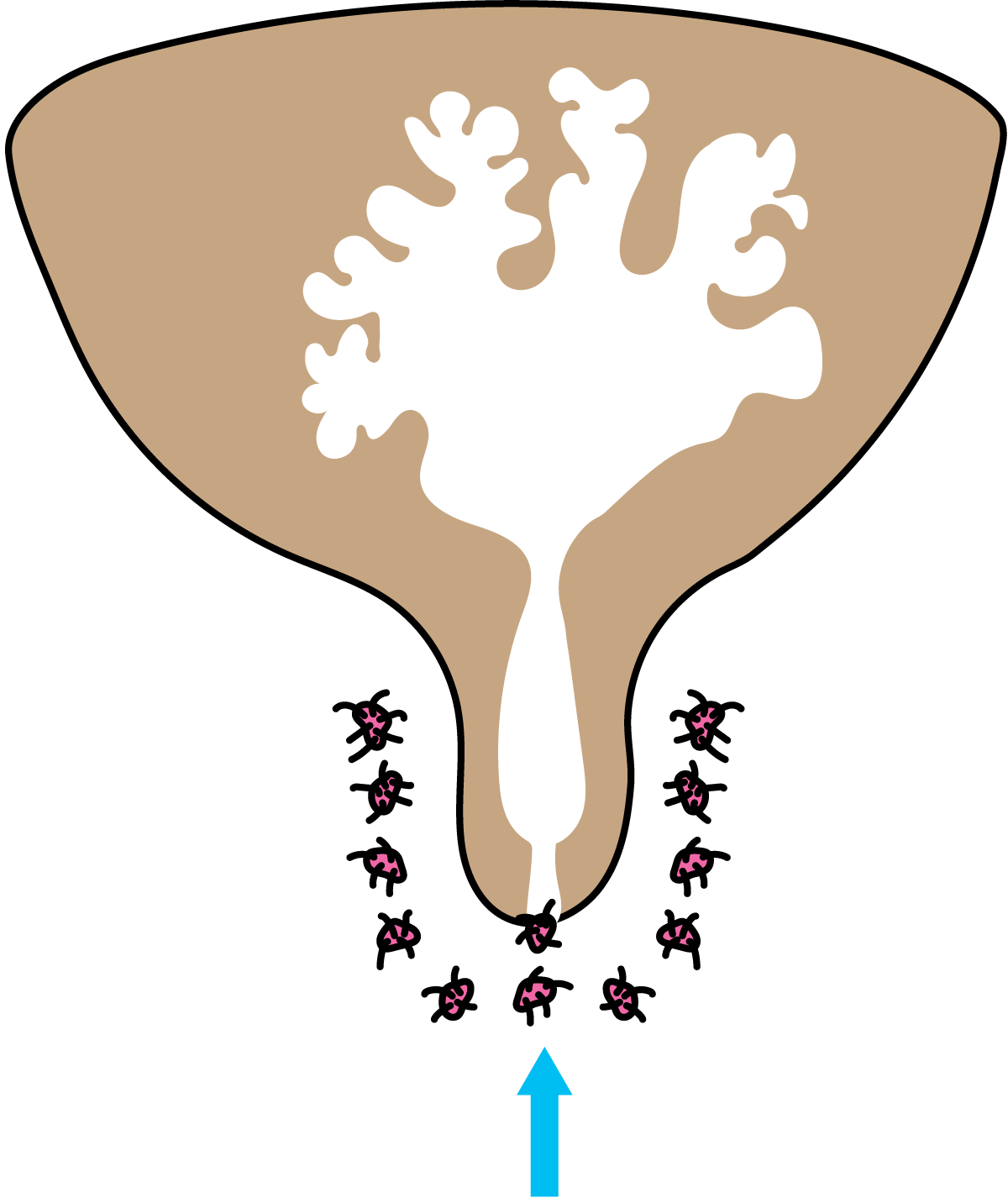
Milk is virtually sterile when it is secreted in the udder. However, even before it leaves the udder, milk is infected by bacteria that enter through the teat canal as shown in Figure 4.15. These bacteria are normally harmless and few in number, up to a few hundred per ml. However, in cases of bacterial udder inflammation (mastitis), the milk is heavily contaminated with bacteria (Figure 4.16) and may even be unfit for consumption.. This condition also causes the cow to suffer.
There are always concentrations of bacteria in the teat canal, but most of them are flushed out at the beginning of milking. It is advisable to collect the first bacteria-rich jets of milk from each teat in a separate vessel with a black cover, as shown in Figure 4.17. Flocculated milk from diseased animals shows up readily against the black background.
Contamination at the farm
In the course of handling at the farm, milk is liable to be contaminated by various microorganisms, mainly bacteria. The degree of contamination and the composition of the bacterial population depend on the cleanliness of the cow’s environment and the cleanliness of the surfaces with which the milk comes into contact, e.g. the pail or milking machine, the strainer, the transport churn or the tank and agitator. Milk-wetted surfaces are usually a much greater source of contamination than the udder.
When cows are milked by hand, bacteria can get into the milk via the milker, the cow, the litter and the ambient air. The magnitude of the influx depends largely on the skill and hygiene consciousness of the milker and the way the cow is managed. Most of these sources of contamination are eliminated in machine milking, but another one is added, namely the milking machine. A very large number of bacteria can enter the milk this way if the milking equipment is not cleaned properly.
The cleanliness of the cow’s teats and udder can have large impact on the spore content of the milk.
Bacteria in raw milk
Milk is very nutritious and is susceptible to contamination and growth of a wide variety of bacteria.
Farm milk may contain anything from a few thousand bacteria per ml, if it comes from a hygienic farm, up to several million if the standard of cleaning, disinfection and chilling is poor. Daily cleaning and disinfection of all milk equipment is therefore the most decisive factor in the bacteriological quality of milk. Under optimal conditions, it should be possible to achieve a bacteria count of less than 20,000 CFU (colony-forming units) per ml. Rapid chilling to below 4 °C contributes greatly to the quality of the milk at the farm. This treatment slows down the growth of the bacteria in the milk, thereby greatly improving its keeping qualities.
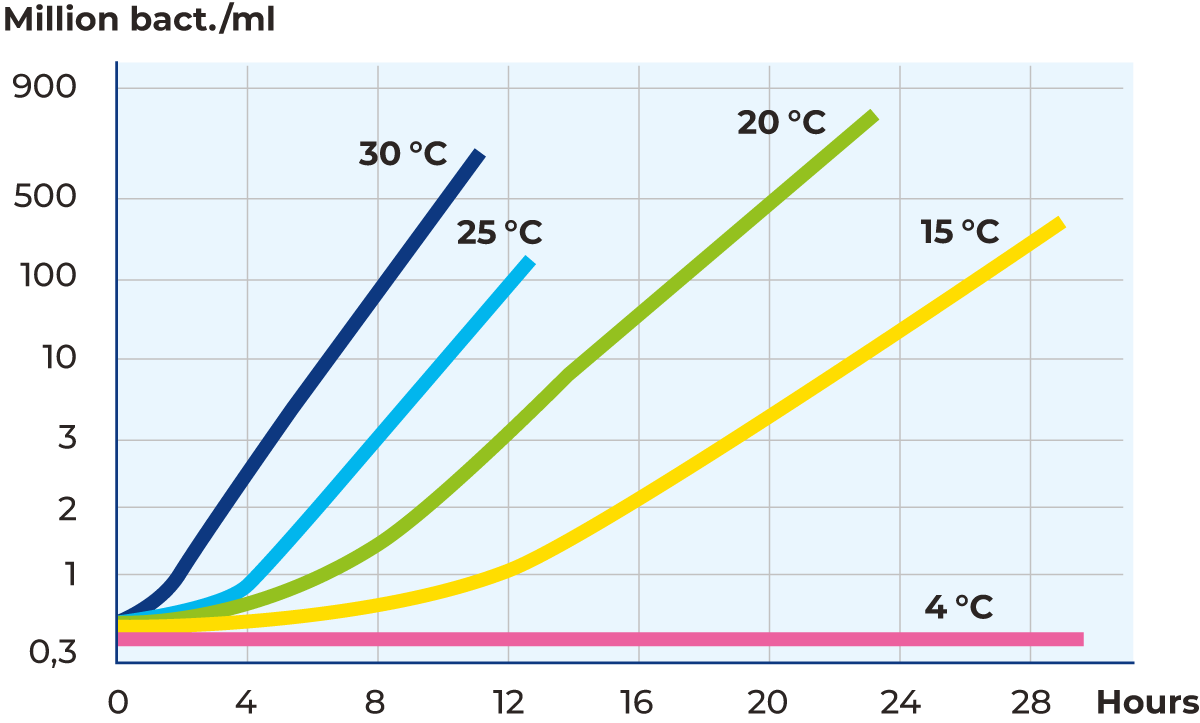
The influence of temperature on bacterial development in raw milk is shown in the graph in Figure 4.18. Starting from 300,000 CFU/ml, the speed of development at higher temperatures and the effect of cooling to 4 °C is striking. Cooling to 4 °C, or even lower to 2 °C, in conjunction with milking makes it possible to deliver milk at two-day intervals provided that the milk container/tanker is insulated.
There are three main sources of contamination: inside the cow, the teats and the udder, and everything the milk comes in contact with. The most common microorganisms in raw milk of good quality (< 20,000 CFU/ml) is restricted to a few groups of bacteria, Table 4.5.
Bacteria in pasteurized milk
HTST (High Temperature Short Time) pasteurization of milk kills heat-sensitive microorganisms. Survivors are spore formers, Corynebacteria and a few other Gram-positive rods and cocci. This relatively heat-resistant flora is called the thermoduric group. In good quality milk, the thermoduric count is usually 200 – 300 CFU/ml.
The occurrence of Gram-negative bacteria in HTST milk depends on water-borne re-infection such as leakage or some other unhygienic condition. As many Gram-negative bacteria can grow at refrigeration temperatures quite well, they spoil the HTST milk in a few days. Therefore, routine laboratory control of Gram-negatives is necessary.
Fungi
Fungi are a group of microorganisms that are frequently found in nature among plants, animals and human beings. Different species of fungi vary a great deal in structure and method of reproduction. Fungi may be round, oval or threadlike. The threads may form a network, visible to the naked eye, in the form of mould on food, for example. Fungi are divided into yeasts and moulds.
Fungi are divided into:
- yeasts
- moulds
Yeasts
Yeasts are single-cell organisms of spherical, elliptical or cylindrical shape. The size of yeast cells varies considerably. Brewer’s yeast, Saccharomyces cerevisiae, has a diameter of 2 – 8 μm and a length of 3 – 15 μm. Some yeast cells of other species may be as large as 100 μm.
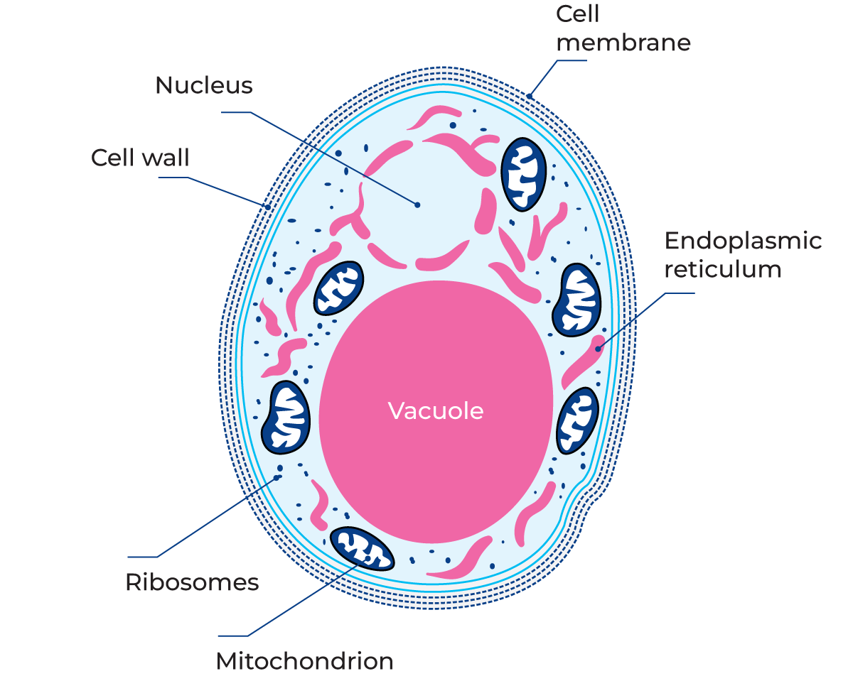
Yeasts, like moulds, have a more complex internal structure than bacteria. They contain cytoplasm and a clearly discernible nucleus surrounded by a nuclear membrane, Figure 4.19. The cell is enclosed by a wall and a cell membrane, which is permeable to nutrients from the outside of the cell and waste products from the inside.
The cell contains a vacuole that serves as storage space for reserve nutrition and for waste products before they are released from the cell. Fat globules and carbohydrate particles are embedded in the cytoplasm. In the cytoplasm there is also a fine network of membranes named endoplasmic reticulum, mitochondria (where energy for cell growth is generated), as well as ribosomes.
Reproduction of yeast
Yeast cells normally reproduce by budding, as shown in Figure 4.20, although other methods of reproduction can also be found. Budding is an asexual process. A small bud develops on the cell wall of the parent cell. The cytoplasm is shared for a while by parent and offspring. Eventually the bud is sealed off from the parent cell by a double wall.
The new cell does not always separate from its parent and may remain attached to it while the latter continues to form new buds. The offspring cell may also form fresh buds of its own. This can result in large clusters of cells attached to each other.
Some types of yeast reproduce sexually, as in Figure 4.21, by forming spores, ascospores and basidiospores (not to be confused with bacterial spores). Two cells fuse together, and the two nuclei also fuse. Following division of the nuclear material, eight ascospores are formed within the cells, each containing a similar set of DNA. When the spores are mature, they are released and germinate, forming new cells, which then reproduce asexually by budding.
Conditions for the growth of yeast
Environment and nutrients
Yeasts have the same need for nutrients as other living organisms. They usually flourish in habitats where sugars are present, such as fruits, flowers, and the bark of trees. A few species are pathogenic to animals and humans. Yeasts are the predominant spoilage organism of fruit juices. Like bacteria, they have a system of intracellular and extracellular enzymes capable of breaking down large molecules in the substrate to manageable size for the metabolism of the cells. In the laboratory, yeasts are cultivated in sugar-rich substrates with pH 5 to 6.
Moisture
Like bacteria, yeasts must have access to water in order to live, but yeasts require less water than bacteria. Some species can grow in media with very low water content, such as honey or jam.
Acidity
Yeast can grow in media with pH values ranging from 3 to 7.5. The optimum pH is usually 5 – 6.
Temperature
Yeast cells do not usually grow at temperatures below the freezing point of water or above about 47 °C. The optimum temperature is normally between 20 and 30 °C. Many genera contain psychrotrophic strains; Candida, Cryptococcus, Rhodotorula and Torulopsis, for example.
Growing cells are normally killed within 5 to 10 minutes at temperatures of 52 – 58 °C. Spores (ascospores) are more resistant but are killed when exposed to 60 – 62 °C for a few minutes.
Oxygen
Yeasts have the ability to grow both in the presence and in the absence of atmospheric oxygen, i.e. yeast are facultatively anaerobic. In the absence of oxygen, yeast breaks down sugar into alcohol and water while, in the presence of oxygen, it breaks down sugar into carbon dioxide and water. Yeast cells grow faster in the presence of oxygen.
Classification of yeasts
Yeasts are divided into three groups, according to their ability to produce spores (ascospores and basidiospores). The strains that form spores belong to the groups Ascomycetes and Basidiomycetes. Those that do not produce spores, but reproduce mainly by budding, belong to the group of Fungi imperfecti.
Importance of yeast
Yeasts are generally undesirable organisms from the dairy point of view, with one exception. Kefir, a Russian cultured product, is fermented with a mixed culture of yeasts and lactic acid bacteria in a grain-shaped aggregate. Otherwise, yeast organisms can cause serious faults in culture dairy products, including cheese and butter. In the brewing, wine, baking and distilling industries, on the other hand, they make a valuable contribution.
Important factors for yeast growth
- nutrients
- moisture
- acidity
- temperature
- oxygen
Yeasts grow best in acid media.
Moulds
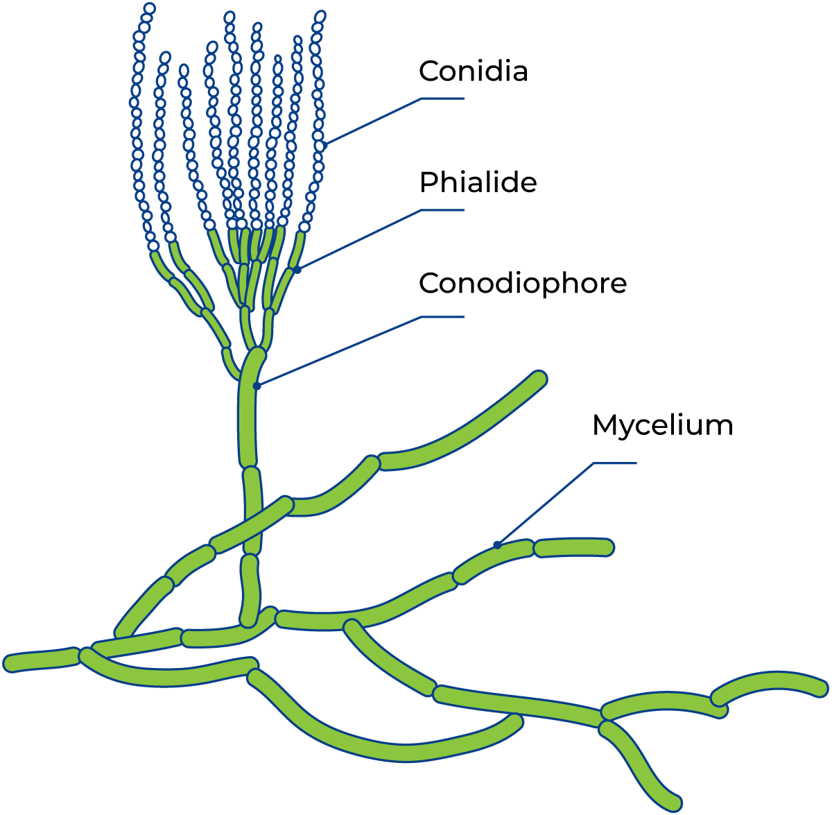
The category of moulds comprises a fairly heterogeneous group of multi-cellular threadlike fungi. The moulds consist of threadlike strands of cells called hyphae. The mass of hyphae that can be seen with the naked eye is called mycelium. The hyphae may or may not have cross-walls between the cells and are usually branched. The hyphae are the vegetative part of the mould, often colourless, and secrete enzymes by which they degrade food (Figure 4.22).
As the mould colony grows, the hyphae and mycelium radiate outwards from the centre.
Reproduction of moulds
Moulds reproduce by means of spores of various types. Both sexual and asexual reproduction may occur in the same species. The asexual spores usually have thick walls and are relatively resistant to desiccation. Some strains have very heat-resistant sexual spores. A mould can remain dormant in spore form for quite a long time.
The asexual spores, conidia, represent the most common method of mould reproduction (figure 4.23), and they are usually produced in enormous numbers. They are very small and light and can be carried by air currents, spreading the mould from place to place. This is a common, everyday occurrence, and often presents a problem in the dairy industry.
Metabolism of moulds
Mould fungi metabolise in much the same way as yeasts. They are well-equipped with enzymes, which they use to break down a variety of organic substances. From the dairy point of view, the action of mould on fat and protein is of particular interest. The growth of mycelium mould is illustrated in Figure 4.24.
Moisture
Moulds can grow on materials with a very low water content and can extract water from moist air.
Water activity (aw)
Moulds are more tolerant to low aw than any other group of microorganisms. Some can tolerate concentrations of sugar and salt with high osmotic pressure, e.g. fruit preserves and sweetened condensed milk.
Oxygen
Moulds normally grow in aerobic conditions. Oxygen is necessary for the formation of conidia, and for the growth of mycelia.
Temperature
The optimum growth temperature for most moulds is between 20 and 30 °C.
Acidity
Moulds can grow in media with pH values from 3 to 8.5. Many species, however, prefer an acid environment, e.g. cheese, yoghurt, citrus fruit and fruit juices.
Importance of moulds in the dairy
As with yeasts, moulds do not survive ordinary pasteurization temperatures, 72 – 74 °C for some 10 – 15 seconds, except moulds with heat-resistant sexual spores. The unwanted presence of these organisms is therefore a sign of re-infection. There are many different families of moulds. Some groups that are of importance in the dairy industry are Penicillium and milk mould, Geotrichum candidum.
Moulds are strictly aerobic organisms (with a few exceptions)
Penicillium
The genus Penicillium is one of the most common types of mould. The spore-forming hyphae of this family are branched at the tip, resembling a brush. Green mould, which occurs widely in nature, belongs to this family. Some species of penicillia play an important part in dairy processes. Their powerful protein- and fat-splitting properties make them the chief agents in the ripening of blue cheese, Camembert, etc. The blue-cheese mould is called Penicillium roqueforti and the Camembert mould, Penicillium camemberti. See Figure 4.23.
Milk mould
The milk mould Geotrichum candidum is on the borderline between yeast and mould. Its reproduction is similar to that of yeast organisms; the outer part of the hyphae is tied off in a process that resembles budding. Its structure is shown in Figure 4.25. The mould occurs on the surface of cultured milk as a fine, white velvety coating. This mould contributes to the ripening of semisoft and soft cheeses. It may cause rancidity in butter.
Moulds on the surfaces of cheese and butter can cause discolouration and also give the product an off flavour. Strict hygiene is necessary in the dairy in order to prevent products from being affected by moulds during processing. Walls and ceilings, for example, must be kept scrupulously clean in order to prevent moulds from settling there.
Bacteriophages
Twort, an English scientist, discovered as early as 1915 that certain cultures of staphylococci were disrupted and broken down. A couple of years later d‘Herelle, a Canadian scientist, after having made similar observations, postulated that the phenomenon was caused by invisible organisms feeding on the bacteria. He called them bacteriophages.
Bacteriophages are thus viruses, i.e. bacterial parasites. By themselves, they can persist, but they cannot grow or replicate except within bacterial cells. They have very specific hosts, e.g. single species or strains of bacteria.
Structure of bacteriophages
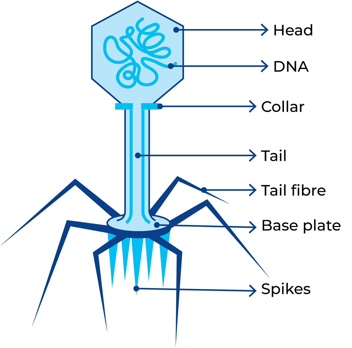
Bacteriophages, or phages, vary considerably in size, and an electron microscope is needed to see them. One of the biggest is the T-phage, which infects Escherichia coli and closely related bacteria. The T-phages have a “head” and a “tail” and a size of 0.03 to 0.3 μm. A schematic drawing of a phage is shown in Figure 4.26.
Reproduction of phages
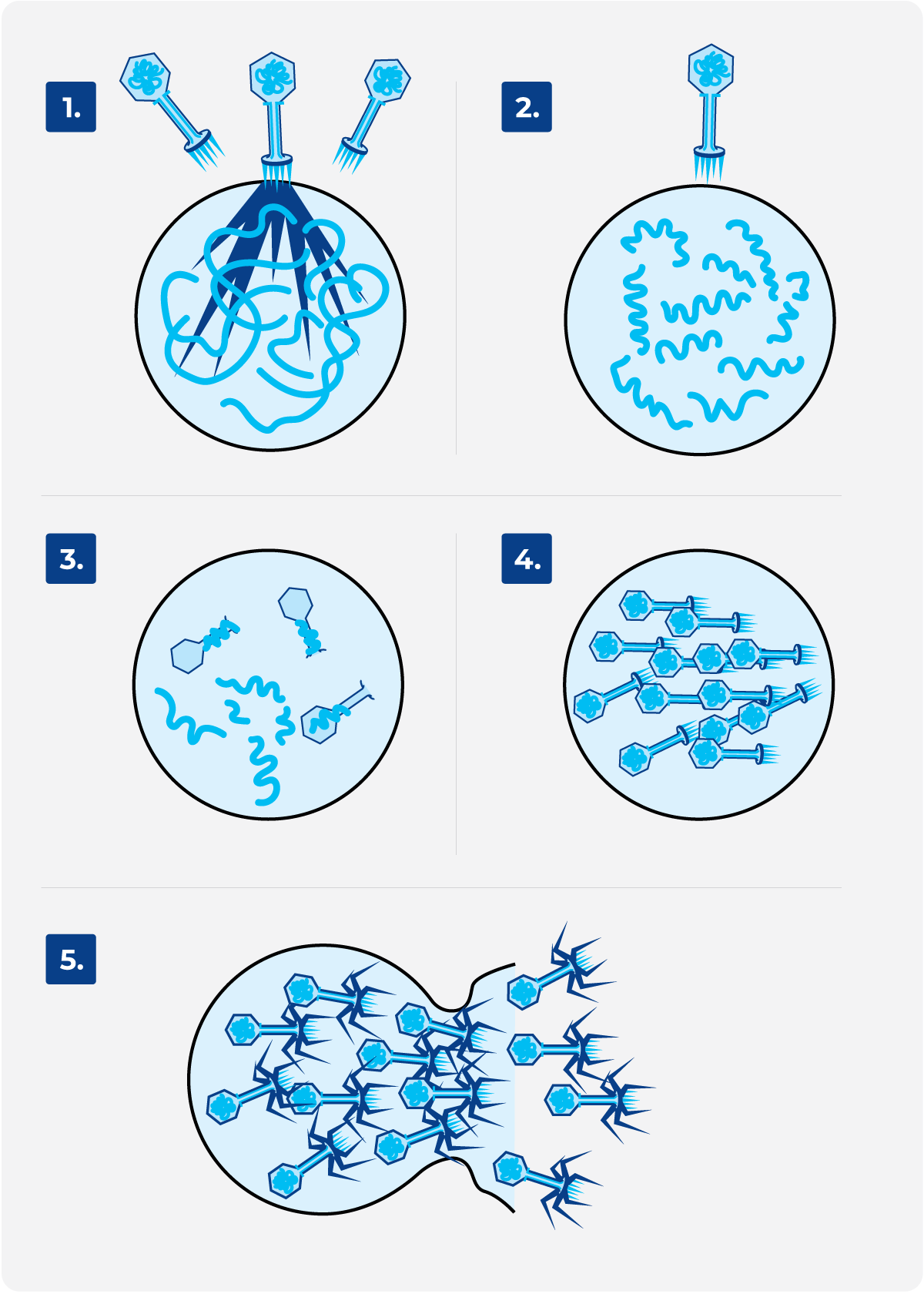
Phages attack actively growing bacteria, within which they can replicate. The bacteria subsequently disintegrate, releasing a group of 10 – 200 phages per bacterium to attack new victims. The scenario is shown in Figure 4.27.
The phage attaches to the surface of its host (1), and the DNA is injected into the cell. The cellular “machinery” then produces new phage DNA and phage proteins (2; 3). The new phages are assembled inside the bacterial cell (4), which is then lysed (5) and the mature phages are released.
Importance of bacteriophages in the dairy
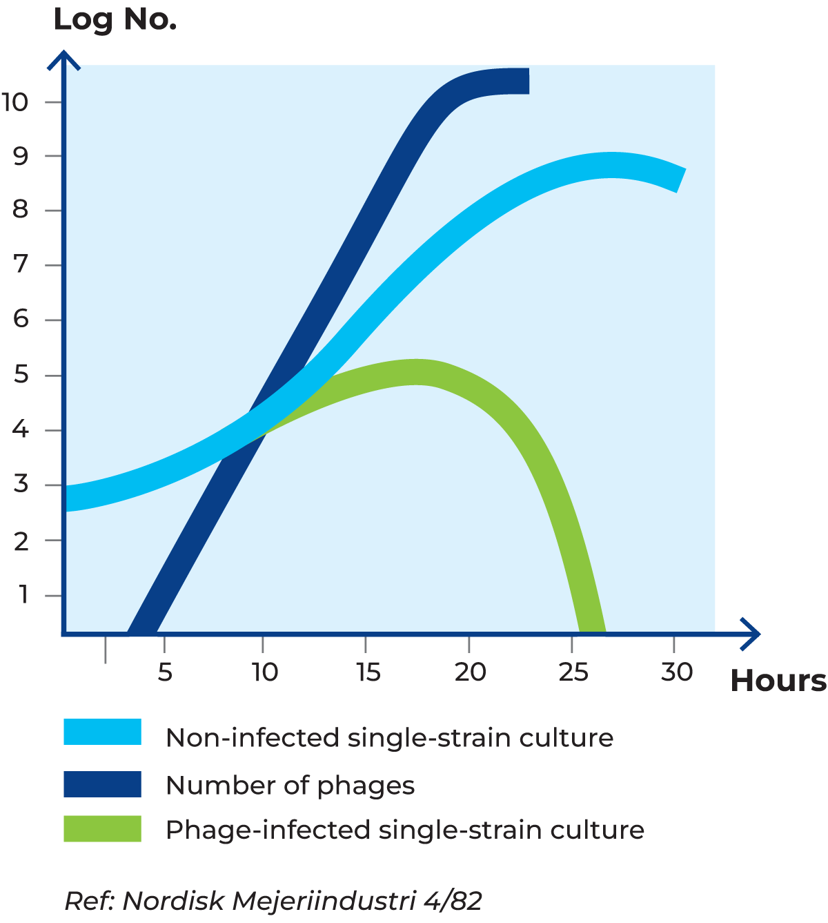
As raw milk is usually contaminated with bacteriophages, it is important that the milk used for starter cultures, generally skim milk, is heated to at least 90 °C for 30 minutes to inactivate the phages. Figure 4.28 shows what will happen if this is not done or if the milk is re-contaminated by phages afterwards. The time it takes for one “non-infected” bacterium to produce four new bacteria is two generations. In the same period, one phage infects one bacterium, which releases 150 new phages in one generation’s time. These phages infect 150 new bacteria and in a further one generation’s time, 22,500 phages are released.
Concluding notes
The great variety of bacteria, yeasts and moulds, and their wide range of activities, are of the utmost importance to life on earth in general, and to humanity in particular.
Microorganisms in soil and water are responsible for degrading available sources of nutrients into forms that plants can assimilate. By doing so, they also perform an indirect service to the animal kingdom, including Man.
Human beings also benefit more directly from microorganisms. Lactic acid-forming microorganisms, for example, can be used to preserve fodder (silage) for livestock. The same principle is applied to the preparation of certain foods such as sauerkraut, green olives and cucumbers.
Microorganisms are of paramount importance in the manufacture of many dairy products, such as yoghurt, cheese and cultured butter. Selection of the right types of microorganisms is an important factor in maximising the quality of these products.
Microorganisms used in the manufacture of dairy products are normally supplied by companies that specialise in developing and propagating them under strictly controlled hygienic conditions. The microorganisms used in the dairy industry are called starter cultures. A starter culture is, for example, a mixture of organisms that form lactic acid by fermenting the lactose in milk. However, it is important that the quality of the starter cultures is preserved after arrival at the dairy by maintaining high standards of hygiene, and that when used, sterile technique is applied in critical steps of the processing chain.
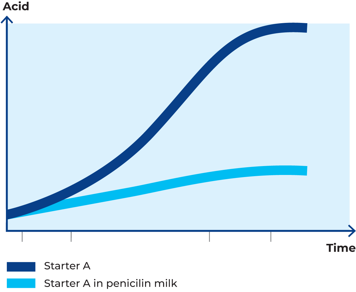
In this context, it should be mentioned that the milk may contain residues of antibiotics emanating from the treatment of cows suffering from mastitis; the most commonly occurring one is penicillin. In spite of regulations saying that milk from cows treated with antibiotics must not be sent to the dairy, you may find sufficiently high levels of antibiotics in bulk tank milk to stop or retard the growth of the starter cultures to be used. Figure 4.29 illustrates the influence of even small residues of penicillin on the most commonly used starter cultures.
It would be a false idealisation of microorganisms to omit to mention that some of them – the pathogenic microorganisms – are regarded as mankind’s worst enemies. The threat against humans is becoming more and more serious due to the use of antibiotics. Some pathogens are today resistant to all antibiotics. It is also true that without the many harmless microorganisms, life would not be possible for humans and many other living organisms.
In most countries, governments have passed laws requiring pasteurization of milk produced at a dairy and intended for consumption. A typical temperature/time combination for pasteurization is 72 °C for 15 – 20 seconds, which kills all pathogens. In order to avoid recontamination, it is of utmost importance that good hygiene practice prevails.
